The extraction of third molars, commonly known as wisdom teeth, is one of the most frequently performed oral surgical procedures. While generally considered routine, the procedure requires a profound understanding of maxillofacial anatomy due to the proximity of these teeth to vital structures. A primary concern for both patients and surgeons is the relationship between the lower wisdom teeth and the inferior alveolar nerve (IAN), a branch of the mandibular nerve.
Understanding the anatomical nuances and the potential for nerve involvement is essential for informed decision-making. This article provides a transparent, clinical examination of the risks associated with nerve proximity during wisdom tooth extraction, the technological advancements used to mitigate these risks, and the surgical protocols employed to ensure patient safety.
Anatomy of the Mandibular Nerve
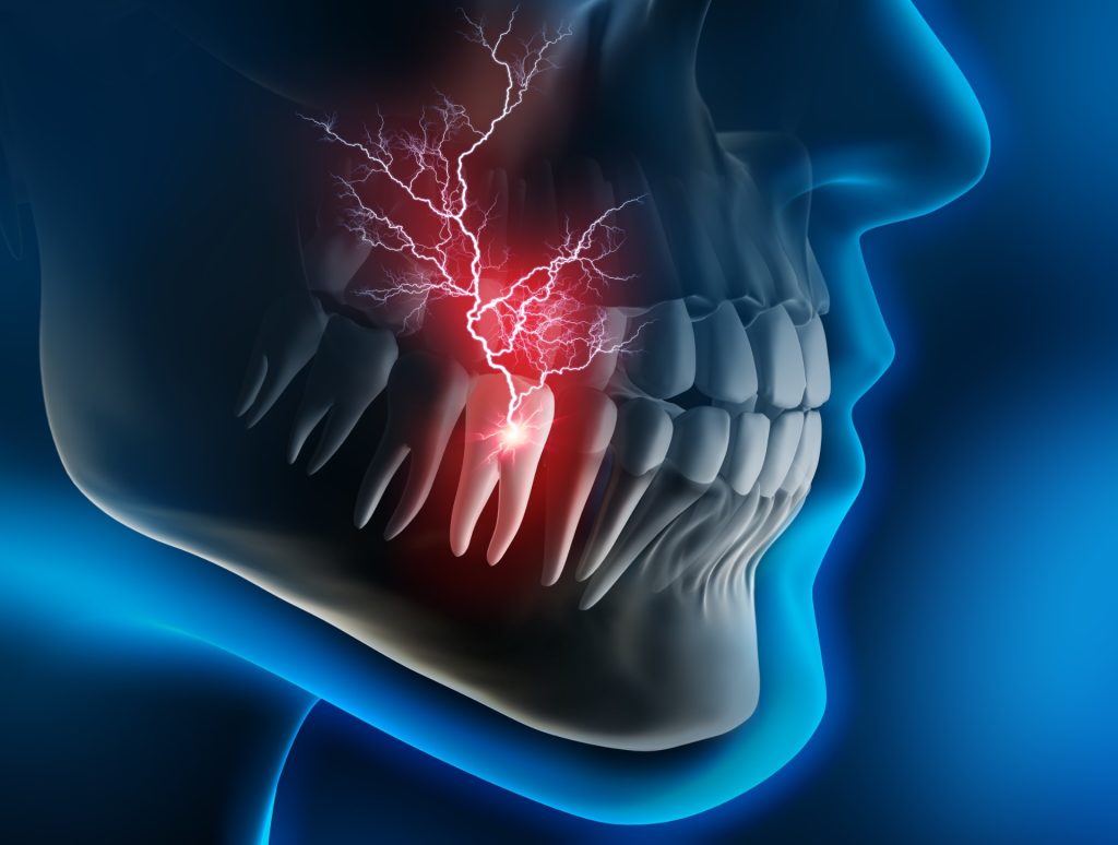
To understand the risks, one must first understand the anatomy. The inferior alveolar nerve runs through a canal within the mandible (lower jawbone). Its primary function is sensory; it supplies sensation to the lower lip, chin, and teeth on that side of the face. It is important to distinguish this from the facial nerve, which controls muscle movement. Injury to the mandibular nerve affects feeling, not the ability to smile or move the face.
During the developmental years, typically late adolescence, wisdom teeth form and begin to erupt. Due to evolutionary changes in jaw size, many individuals lack sufficient space for these teeth to emerge properly. This often results in wisdom teeth removal becoming a medical necessity to prevent infection, crowding, or damage to adjacent molars. However, as the roots of these teeth develop, they grow downward, often coming into close contact with the mandibular canal where the nerve resides.
Classifying the Risk: Root Proximity and Impaction
Not all wisdom teeth pose the same level of risk. The potential for nerve interaction is largely dictated by the position and angulation of the tooth, as well as the length and curvature of the roots.
Vertical and Horizontal Impaction
When a tooth is impacted, meaning it is trapped beneath the gum or bone, the surgical approach becomes more complex. In cases of vertical impaction, the tooth may sit directly on top of the nerve canal. In horizontal impactions, the crown of the tooth pushes against the adjacent molar, while the roots may project towards the nerve. Understanding dental health complications from impacted wisdom teeth helps clarify why leaving high-risk teeth in place is not always a viable option, even when nerve proximity is a factor.
Anatomical Variations
In some patients, the roots of the wisdom teeth may physically wrap around the nerve or press against the canal, causing a compression of the neurovascular bundle. In rare instances, the nerve may even pass through the roots of the tooth. Identifying these specific anatomical configurations is crucial prior to any surgical intervention.
Advanced Imaging and Diagnostic Precision
Historically, surgeons relied on two-dimensional panoramic X-rays to assess wisdom teeth. while useful for a general overview, 2D imaging has limitations in determining the depth and exact spatial relationship between the tooth root and the nerve.
Today, the standard of care for high-risk cases involves the use of cone beam computed tomography (CBCT). This 3D imaging technology allows the qualified oral surgeon to visualize the anatomy from every angle. By slicing the image digitally, the surgeon can see exactly where the nerve lies relative to the root structure—whether it is buccal (cheek side), lingual (tongue side), or inferior (below) the tooth.
This level of computer-assisted surgery planning significantly reduces the element of surprise during the procedure. It allows the surgical team to map out a precise extraction path that avoids the nerve or to decide on alternative surgical techniques if the risk of full extraction is deemed too high.
Surgical Techniques to Mitigate Nerve Injury
When diagnostic imaging confirms a high risk of nerve injury, specialized surgical techniques are employed.
Standard Extraction with Sectioning
In many cases, the tooth is sectioned (cut) into smaller pieces before removal. This minimizes the amount of bone that needs to be removed and allows the surgeon to lift the tooth parts away from the nerve gently, rather than pulling the tooth out in one piece, which could drag the root against the nerve bundle.
Coronectomy: A Strategic Alternative
For patients where the roots are intimately entangled with the nerve, a procedure known as a coronectomy may be recommended. In this procedure, the surgeon removes only the crown of the tooth (the part causing infection or crowding) while intentionally leaving the roots and the nerve undisturbed within the jawbone.
The remaining roots generally heal over with bone and do not cause future problems. This approach virtually eliminates the risk of direct injury to the inferior alveolar nerve during extraction. It is a prime example of how experience matters for complex surgery, as determining candidacy for coronectomy requires high-level clinical judgment.
Understanding Potential Complications
Despite the most advanced imaging and surgical techniques, the risk of nerve involvement cannot be reduced to zero in anatomically complex cases. It is vital for patients to understand the clinical terminology associated with nerve risks.
* Paresthesia: This refers to an altered sensation, typically described as numbness or a “pins and needles” feeling in the lip, chin, or tongue. This is the most common presentation of nerve injury and is usually temporary, resolving as the nerve heals from inflammation or compression.
* Dysesthesia: This involves an abnormal, sometimes unpleasant or painful sensation.
* Anesthesia: This refers to total numbness in the affected area.
The vast majority of nerve injuries related to wisdom tooth extraction are transient. The nerve tissue may be bruised or compressed by the swelling associated with surgery, leading to temporary numbness that resolves over weeks or months. Permanent nerve damage is a rare complication, occurring in a very small percentage of cases.
Post-Operative Monitoring and Recovery
The recovery period is critical for monitoring nerve function. Patients are provided with detailed home care instructions after oral surgery. If a patient experiences numbness that persists beyond the expected duration of the local anesthetic (typically 24 hours), they are instructed to contact their surgeon immediately.
Early intervention and mapping of the sensory deficit are part of the follow-up protocol. Surgeons may prescribe anti-inflammatory medications to reduce pressure on the nerve or recommend neurosensory testing to track improvement. Following tips for a smooth recovery can also help reduce general inflammation, creating a better environment for healing.
The Role of Specialized Care
The extraction of wisdom teeth near the mandibular nerve is not a simple dental procedure; it is a complex surgery requiring a deep understanding of neuroanatomy. This is why many general dentists refer these cases to Oral and Maxillofacial Surgeons.
Surgeons with dual degrees (MD and DDS) and board certification possess extensive training in managing complications and performing delicate dissection around nerves. Their training includes hospital-based rotations in anesthesia, general surgery, and neurosurgery, providing a comprehensive medical background. For patients concerned about anxiety or the complexity of the procedure, options for sedation and general anesthesia are available and administered with the highest safety standards.
Navigating Complex Anatomical Structures
The proximity of wisdom teeth to the mandibular nerve presents a calculated risk that can be managed effectively through science, technology, and surgical skill. While the possibility of nerve interaction exists, the use of 3D CBCT imaging, strategic surgical planning such as coronectomy, and the expertise of board-certified specialists significantly mitigate these risks.
Patients are encouraged to ask questions regarding their specific anatomy during consultations. A transparent discussion about risk factors, combined with a clear surgical plan, empowers patients to proceed with treatment confidently, prioritizing their long-term oral health and overall well-being.
About Head & Neck Surgical Associates
Head & Neck Surgical Associates (HNSA) is the Northwest’s most specialized surgical practice, uniquely staffed by dual-degreed, board-certified physicians trained in both medicine and dentistry. Based in Portland, Oregon, their team offers expert care in head and neck surgery, oral and maxillofacial procedures, and facial cosmetic surgery—backed by advanced fellowships and decades of focused experience. Patients trust HNSA for precise diagnoses, complex surgical treatment, and comprehensive care from true leaders in their field.
For more information or to schedule a consultation, visit our website or connect with us on social media: